Shockwaves

Shock waves are acoustic waves with extremely high energy peaks, such as those produced in the atmosphere by explosive phenomena like lightning strikes or sonic explosions. A shock wave differs from ultrasound in its extremely wide pressure amplitude. What’s more, ultrasound usually consists of a periodic oscillation, whereas a shockwave is a single pulse.
Extracorporeal shockwave therapy (ESWT) is the application of shockwaves in medicine.
When applied to damaged tissue, pressure waves stimulate metabolic reactions. It’s clinically proven:
- Reduces pain felt by nerve fibers
- Increased blood flow to surrounding soft tissue
- Stem cell activation initiates the healing process
Evolution of shockwave therapy
The term “shockwave therapy” refers to mechanical pressure impulses that spread like a wave through the human body. In 1980, the shock wave method was first used to disintegrate stones in a patient’s kidneys(Journal of Urology, 1982).
Therapeutic effect of shockwaves
Extracorporeal acoustic wave therapy (unlike lithotripsy) is not used to disintegrate tissue, but to induce microscopic extracellular and interstitial biological effects, including tissue regeneration. In modern pain therapy, acoustic wave energy is conducted from the point of origin, which is the acoustic wave generator (via a coupling gel), to the areas of the body experiencing pain. Here, its curative capacity is applied.
After 4 or 5 sessions, over 80% of patients report no more pain or a very marked reduction in pain.
Benefits for the patient:
- Wide range of applications
- Anesthetic-free, non-traumatic treatment
- High recovery rate
- Quality of life and mobility restored
- Affordable therapy
- Motivation by visual assessment process with VAS result
- 80% success rate
- Short processing time: approx. 10 minutes
- On average 6-8 treatment sessions required
- Realistic alternative to surgery
The 2 types of shockwave
Radial shockwaves have been used for many years to treat tendinopathies. Your physiotherapy practice in Bascharage also uses focal shockwaves, which are much more precise and effective for many pathologies. What’s more, not all radial shockwaves are created equal, and this can be one of the reasons for the success or failure of a treatment.
Radial shock waves
Ballistic shockwave generation
Compressed air accelerates a projectile, which strikes a fixed applicator at high speed (up to 90 km/h). The kinetic energy is converted into a shockwave that is released through the skin into the targeted tissue. These shockwaves propagate radially across large treatment areas.

Pressure characteristics of radial shock waves
- The shock wave begins with a compression phase that propagates shear waves through the tissue.
- This is followed by a phase of depression or tension that generates cavitation bubbles.
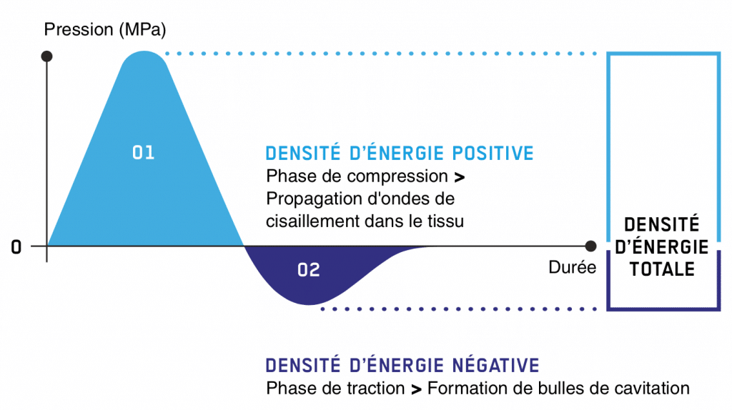
The energy flux density (ED or EFD) is represented by the square area below the pressure curve.
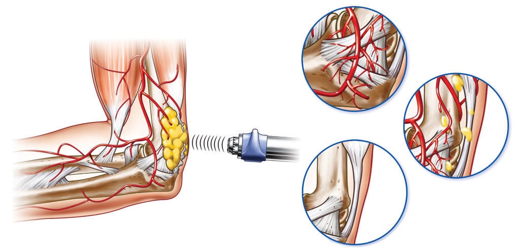
Visualization of radial shock waves

Focal shock waves
Piezoceramic shockwave generation
A high voltage is applied to 1,000 piezoceramic crystals, generating pressure waves. They penetrate the skin and pass through the tissue, concentrating on a cigar-shaped volume.

Pressure characteristics of radial shock waves
- The compression phase of focused extracorporeal shockwaves is usually shorter than that of radial extracorporeal shockwaves, and the maximum pressure P+ is usually higher than in radial ESWT.
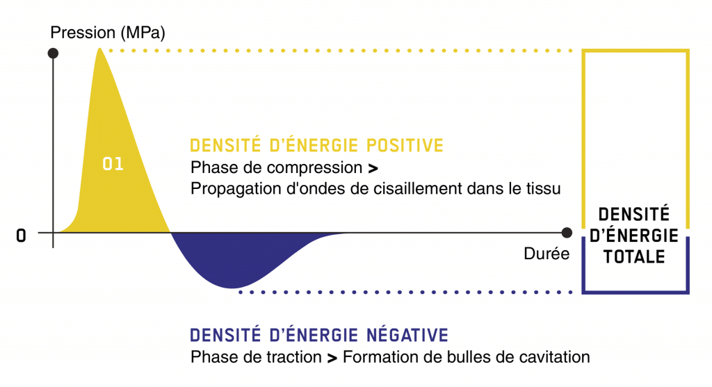 Focused ESWT and radial ESWT can both achieve an ED+ of 0.4 mJ/mm2, a value that has been proven for almost all ESWT indications on the musculoskeletal system and skin.
Visualization of focal shock waves

The 2 major impacts of shockwave therapy
Tissue repair and cell growth
Inward growth of neovascularization :
The blood flow of nutrients is necessary to start and maintain the repair processes of the damaged tissue structure. The application of acoustic waves creates capillary microruptures in tendon and bone and significantly increases the expression of growth indicators such as eNOS, VEGF, PCNA and BMP.
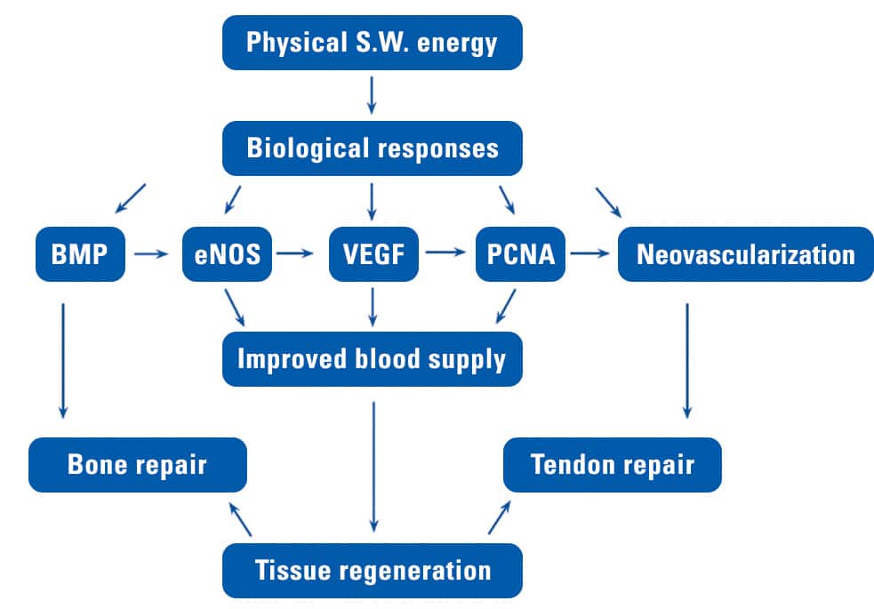
Both processes stimulate the growth and remodelling of new arterioles. The new blood vessels will improve blood supply and oxygenation, resulting in faster healing of tendon and bone.
Reversing chronic inflammation:
Chronic inflammation occurs when the inflammation reaction is not completely stopped. It can damage healthy parts of the body and lead to chronic pain. The activity of mast cells, involved in the inflammatory process, can be enhanced by insinuating acoustic waves. Mast cell activation can be followed by the synthesis of chemokines and cytokines. Unblocking pro-inflammatory mixtures, if necessary, can help restore normal healing and regenerative processes.
Collagen stimulation :
The production of sufficient quantities of collagen is a necessary precondition for the tissue repair process. Shockwave therapy accelerates procollagen synthesis. The newly created collagen fibers are imposed in a longitudinal structure. These newly-formed tendon fibers are denser and harder.
Analgesia and mobility rehabilitation
Dispersion of pain mediator Substance P :
Substance P is a neurotransmitter that mediates pain information via C-fibres. This neuropeptide is generally associated with chronic, persistent and intense pain. It is used to relay pain messages to the central nervous system. Reducing the concentration of substance P reduces the stimulation of afferent nociceptive fibers, thereby reducing pain. Reduced substance P, histamines and other nociceptive metabolites also help inhibit the development of inflammatory edema.
Dissolution of calcified fibroblasts :
Calcium deposits are most often the result of micro-tears or other tendon trauma. Acoustic waves break up existing calcifications. Acoustic wave therapy initiates biochemical decalcification of the toothpaste-like calcium deposit and treats the tendon. Calcium granules are removed by the lymphatic system.
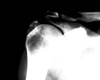 Pre-treatment x-ray images.
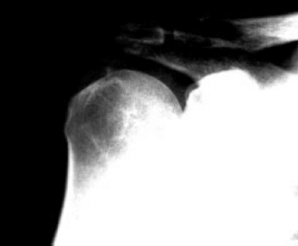 X-ray images after 3 shockwave sessions
Trigger point relief:
Trigger points are the main cause of pain in the back, neck, shoulders and extremities. They are associated with palpable nodules in taut groups of muscle fibers, and their sarcomeres are extremely contracted. Dysfunctional sarcomeres contract so hard that they begin to cut off their own blood supply. This causes a deposit of waste products which in turn irritates the sensory nerve endings, causing even more contractions. This vicious circle is referred to as a “metabolic crisis”. Although the precise medical effects of the acoustic wave are still unclear, it can be argued that the acoustic energy supplied unlocks the “calcium pump”. This reverses the metabolic crisis into myofilaments and relieves these trigger points.
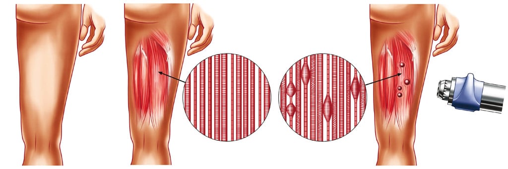
Acoustic waves relieve extremely contracted sarcomeres in trigger points.
Shockwave applications
Extracorporeal shockwave therapy is most frequently used in physiotherapy, orthopedics and sports medicine.
- Sore shoulders
- Tennis player’s lateral epicondylalgia/elbow
- Medial epicondylalgia/golfer’s elbow
- Lower back pain
- Achilles tendon pain
- Tendinopathies and chronic enthesopathies in general
- Trigger point treatment
- Dupuytren’s disease
- Ledderhose disease
- A frozen shoulder
- A periarthritis humeroscapularis
- Arthrotic changes: secondary symptoms
- Calcaneal spur/plantar fasciitis
- Shin splints/Anterior tibial syndrome
- Patellar tendinopathy
- Achillodynia
- Myofascial pain syndrome
- Muscular distension
- Prolonged treatment of joint distortion
- Groin pain
- Hip pain
- Lower back pain
- Achillodynia
- Iliotibial band syndrome
Shockwave therapy is the perfect treatment method for all complex chronic and musculoskeletal conditions.
New indications for shockwave treatment in sports medicine
This therapy can be used for chronic and acute soft-tissue disorders in sports medicine. The latest results prove the effectiveness of shockwaves in healing muscular tension, reducing recovery times and enabling a return to full sporting endurance.
Sports medicine indications for shock waves are :
- Healing acute muscular tension
- Support for muscle regeneration
- Acute muscle spasms
- Jumper’s knee
- Epicondylitis
- Shoulder pain
- Groin pain
- Achillodynia
- …
In the case of acute muscle injury, shockwave therapy is part of my standard therapeutic procedure. I’ve managed to use them for all my patients, including some of the world’s best tennis players, including Tomas Berdych (2010 Wimbledon finalist) and other members of the Czech Davis Cup team.
I had a case of a badminton player with bilateral achillodynia. After the fourth treatment there was still some pain, but a month after the last treatment the pain had completely disappeared.
This therapy is an innovative method for treating the musculoskeletal system in sports physiotherapy. It’s non-invasive, safe and effective. In around 10 days, shockwave therapy can reduce pain in 84% of cases.
This therapy is an approach to be favored in a complex course of treatment focused on optimizing inflammation and reducing the healing interval. Early activation of the injured muscle and stimulation with shockwaves play a crucial role in faster muscle recovery.
Cryotherapy combined with shock waves
Cryotherapy combined with shock waves is a must!
Shockwave therapy can be uncomfortable and even painful. There is also a risk of bruising. The use of cryotherapy BEFORE shockwave therapy is beneficial:
- Its analgesic effect makes the treatment totally painless and offers greater comfort for the patient. In this way, it is possible to use appropriate parameters to improve the effectiveness of the therapy.
- Vasomotor effects reduce the risk of contusion caused by shockwave therapy.
- Escape allows you to improve the healing process and increase the effectiveness of therapy.
Cryotherapy (thermal shock), combined with shock waves, improves patient comfort, makes therapy gentler and increases effectiveness. Cryotherapy is the therapeutic application of cold, which can be obtained in a variety of ways (ice, cold water, damp cloth, methyl chloride, carbon dioxide, etc.).